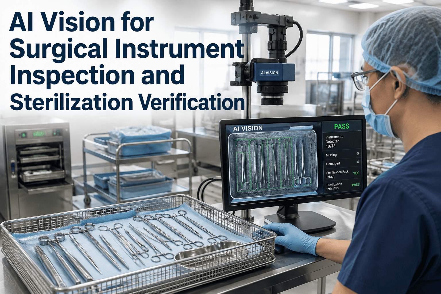
A surgical tray is checked by human eyes under fluorescent light, often during the busiest hour of a shift, and that single visual pass is frequently the only thing standing between a damaged clamp or a speck of retained tissue and a patient on the table. Sterile processing departments report that the vast majority of instrument-related errors trace back to visualization failures rather than the sterilization cycle itself, because a scratched jaw, a cracked insulation coating, or a hairline lumen crack is easy to miss under time pressure. AI vision cameras are now being placed at the inspection bench itself, checking every instrument for damage, residual bioburden, and tray completeness before it ever reaches sterilization. To see how this fits your sterile processing workflow, book a demo.
HEALTHCARE VISION · STERILE PROCESSING · INSTRUMENT INTEGRITY
The Inspection Step Sterile Processing Can't Afford to Get Wrong
Every instrument that reaches an operating table has already passed through a visual check that decides whether it is safe to use. AI vision turns that single, fatigue-prone check into a consistent, documented, camera-verified step — before sterilization, not after a complication.
FROM OPERATING ROOM BACK TO OPERATING ROOM
Tracing an Instrument's Path Through Sterile Processing
An instrument that leaves surgery contaminated has to pass through several handoffs before it returns to a tray, and the inspection step sits at the point where damage, debris, and incomplete cleaning are the easiest to catch and the easiest to miss.
01
Decontamination and Cleaning
Instruments are manually or automatically washed to remove blood, tissue, and soil, but lumens, hinges, and box-locks can trap residue that a wash cycle alone does not always clear.
02
Visual Inspection Bench
A technician checks each instrument for cleanliness, function, and damage under a lighted magnifier, a task that studies link to the largest share of downstream instrument errors.
03
Tray Assembly
Instruments are matched to a count sheet and arranged in trays, where a missing or substituted instrument often is not caught until the tray is opened in the operating room.
04
Sterilization
Sterilization kills microorganisms but cannot compensate for instruments that entered the cycle with retained bioburden, since organic residue can shield microbes from steam or gas penetration.
05
Back to the Operating Room
A compromised instrument that reaches this point has already passed every check the department has, which is why the inspection step upstream carries so much weight.
91%
Of cumulative instrument error risk in one multi-hospital study was tied to tasks performed in the sterile processing environment
88.6%
Of observed surgical instrument errors in a pediatric OR study stemmed from visualization failures — missing, broken, or contaminated instruments
94%
Of inspected lumened instruments in a published pilot study showed visible debris or discoloration inside the lumen after standard reprocessing
1.18%
Package-level error rate recorded across nearly 34,000 tracked instrument packages in a hospital sterile supply department study
WHAT THE CAMERA IS TRAINED TO CATCH
Three Categories of Instrument Failure, One Inspection Pass
Manual inspection asks one technician to evaluate cleanliness, function, and completeness simultaneously, under time pressure, for every instrument on every tray. AI vision splits that same evaluation into three parallel, camera-driven checks that do not degrade as the shift goes on.
Physical Damage and Wear
High-resolution imaging flags corrosion, pitting, bent jaws, chipped insulation coating on electrosurgical instruments, and hairline cracks near hinges and box-locks that are easy to miss under standard bench lighting.
Residual Bioburden and Debris
Vision models trained on cleaned-and-sterilized instrument imagery are tuned to spot staining, discoloration, and visible soil in cavities and lumens, the same defect class that published lumen-inspection studies found in the overwhelming majority of instruments examined.
Assembly and Count Completeness
Fine-grained, model-specific recognition distinguishes near-identical instrument variants and cross-checks a tray against its count sheet, catching missing, mismatched, or wrong-specification instruments before the tray is sealed for sterilization.
Bring camera-verified inspection to your sterile processing bench
iFactory configures instrument-level AI vision around your own instrument sets, your own count sheets, and your own inspection bench layout — not a generic industrial defect model repurposed for healthcare.
MANUAL VS. CAMERA-VERIFIED INSPECTION
What Changes at the Inspection Bench
The inspection task itself does not change — cleanliness, function, and completeness still have to be verified before sterilization. What changes is whether that verification depends on one technician's attention holding steady for an entire shift, or on a consistent camera check applied to every instrument, every time.
| Inspection Factor |
Manual Visual Check |
AI Vision Inspection |
| Consistency across a shift |
Catch rate declines with fatigue and time pressure |
Same detection standard applied to the first and the last instrument |
| Lumen and cavity visibility |
Limited without a borescope, often skipped under time pressure |
Targeted imaging for cavities and lumens flagged for closer review |
| Instrument identification |
Relies on technician memory for near-identical variants |
Model-specific recognition trained on the exact instrument set in use |
| Documentation |
Manual log entry, often summarized rather than per-instrument |
Per-instrument image and result logged automatically for traceability |
| Tray completeness check |
Visual count against a paper or digital sheet |
Automated cross-check between imaged instruments and the count sheet |
HOW THE INSPECTION STATION FITS IN
Where AI Vision Sits in the Sterile Processing Workflow
The inspection camera is positioned at the existing bench, immediately after decontamination and before tray assembly, so the workflow technicians already follow does not need to be redesigned around the new inspection layer.
1
Instrument Placed Under the Camera
A technician places a cleaned instrument on the inspection surface as part of the normal workflow, with no separate scanning step added to the process.
2
Multi-Angle Image Capture
Calibrated lighting and multiple camera angles capture the instrument surface, joints, and, for lumened instruments, the cavity opening for closer analysis.
3
Damage, Bioburden, and ID Analysis
The vision model runs damage detection, residue detection, and instrument identification against the reference set in parallel, rather than as sequential manual checks.
4
Pass, Flag, or Recleaning Alert
A cleared instrument proceeds to tray assembly, while a flagged instrument is routed back for recleaning or removed from service, with the reason recorded against that instrument's record.
5
Traceable Record Attached to the Tray
Every inspected instrument's result is logged and linked to the tray it was assembled into, producing a documented inspection trail rather than a single technician's sign-off.
WHY VISUAL INSPECTION ALONE OFTEN ISN'T ENOUGH
The Gap Between a Passed Inspection and a Sterile Instrument
Regulatory guidance for reusable medical devices treats visual inspection as a required step for every device, and as the only step for lower-risk devices, which places a significant amount of weight on a check that depends entirely on human attention and lighting conditions at the bench. Higher-risk critical and semi-critical instruments carry additional residual-marker testing requirements precisely because visual inspection by itself has known limits, particularly for cavities and lumens that are not fully visible without magnification or a borescope.
Published research on sterilization wrap and instrument inspection has raised the same concern from a different angle: a visual pass at the bench does not always correlate with what is actually happening inside a lumen or underneath a hinge, since organic soil can sit in places a standard inspection light does not reach. This is the gap camera-based inspection is built to close, not by replacing the judgment of sterile processing staff, but by giving every instrument the same close, consistent, documented look that a rushed manual pass cannot guarantee on every tray, every shift.
BEFORE A DEPLOYMENT CONVERSATION
What Sterile Processing Leadership Should Have Ready
Departments that move fastest from evaluation to a working pilot generally arrive with a working knowledge of their own instrument mix and their current inspection process, rather than starting that conversation from scratch during the first vendor call.
1
Instrument Set Inventory
A list of the instrument types, models, and manufacturers most commonly processed, since fine-grained recognition is trained against the specific sets in use.
2
Current Error and Rework Data
Any existing tracking of missing, damaged, or recleaned instruments, which becomes the baseline a pilot is measured against.
3
Bench Layout and Lighting
The physical layout of the inspection station, since camera placement and lighting calibration are configured to the existing bench rather than requiring a redesigned workspace.
4
High-Risk Instrument Priorities
Which instrument categories — lumened, hinged, or electrosurgical — carry the highest current concern, so the pilot scope reflects where the risk is actually concentrated.
Start with a baseline review of your inspection bench
A short working session maps your instrument mix, your current error tracking, and your bench layout against what a pilot deployment would look like for your sterile processing department.
FREQUENTLY ASKED QUESTIONS
What Sterile Processing Teams Ask Before Adopting AI Vision
Does AI vision inspection replace the visual inspection step required by reprocessing guidelines?
No. Visual inspection remains a required part of the reprocessing workflow for every device category, and camera-based inspection is built to strengthen that exact step rather than remove it from the process. The system performs the same visual check a technician performs, but applies it consistently to every instrument regardless of shift length or workload, and produces a documented image-based record that a purely manual check does not generate on its own. Sterile processing leadership stays in control of acceptance criteria and escalation decisions throughout.
Contact our support team to review how this fits alongside your existing reprocessing validation documentation.
Can the system actually see inside lumens and cavities, or only external surfaces?
Lumened instruments are one of the harder inspection problems in sterile processing, since published pilot studies inspecting instrument lumens with cameras and borescopes have found visible debris or discoloration in a large majority of examined units. Camera-based inspection targets cavity openings and, where instrument design allows, internal imaging to flag instruments that need borescope follow-up or recleaning, rather than treating every lumened instrument as a black box that passes on external appearance alone.
Book a demo to see how lumen and cavity checks are configured for your specific instrument sets.
How does the system tell apart nearly identical instrument variants during tray assembly?
Standard object detection often struggles with instruments that share the same general shape but differ by manufacturer, size, or model, which is exactly where tray assembly errors involving wrong-specification instruments tend to originate. Fine-grained, model-specific recognition is trained on the exact instrument catalog a department uses, rather than a broad category classifier, so a size mismatch or a substituted variant is flagged against the count sheet before the tray is sealed and sent to sterilization.
Contact our support team to walk through how your instrument catalog would be modeled.
What happens when the system flags an instrument — does it stop the whole tray?
A flagged instrument is routed for recleaning, closer manual review, or removal from service depending on the flag type, while the rest of the tray continues through the normal workflow without an unnecessary full-tray hold. Every flag is logged against that specific instrument along with the image that triggered it, giving the reviewing technician the same visual evidence the system used rather than an unexplained rejection. This keeps the department's throughput close to normal while still catching the individual instruments that need attention.
Book a demo to see the flag-and-review workflow in action.
How long does it take to get an inspection station like this running in our department?
Timelines depend on instrument catalog size and bench configuration, but a scoped pilot on a limited instrument set and a single inspection bench is generally the fastest way to validate detection accuracy against your department's own baseline before considering a wider rollout. Because the system is positioned at the existing bench rather than requiring new workstations, most of the pilot timeline goes toward model calibration against your specific instrument sets rather than physical installation.
Contact our support team to scope a pilot timeline for your department's instrument mix.
EVERY INSTRUMENT, EVERY TRAY, THE SAME STANDARD
Bring Consistent, Documented Inspection to Your Sterile Processing Bench
The inspection step decides what reaches the operating table. iFactory configures camera-based inspection around your instrument sets, your count sheets, and your bench, so that standard holds on every tray, every shift.